Le poumon gauche d'un covidé , au rayon X
De : https://www.medscape.com/viewarticle/962916?src=soc_tw_211116_mscpedt_news_mdscp_xray&faf=1
Via ZAZA40

Traduction automatique, non revue ni corrigée : https://translate.google.com/translate?hl=fr&sl=de&tl=fr&u=https://lezarceleurs.blogspot.com/2021/11/le-poumon-gauche-dun-covide.html
New X-Ray Technique Shows COVID-19 Lung in Unprecedented Detail
A recent article published in Nature Methods highlights how hierarchical phase-contrast tomography (HiP-CT), an x-ray phase propagation technique that uses spatial coherence to conduct three-dimensional scans of organs ex vivo, may offer clinicians greater insights into disease processes.
"It is not a clinical technique as such," said Claire Walsh PhD, a biophysicist and senior research fellow at the Center for Advanced Biomedical Imaging, University College London, London, United Kingdom, and one of the authors of the article. She stressed that HiP-CT is used ex vivo.
"This technology uses x-rays from a fourth-generation x-ray source, the European Synchrotron Radiation Facility's Extremely Brilliant Source. It is an incredibly bright x-ray source," said Walsh in an interview with Medscape Medical News. She said synchrotron x-ray tomography provides a much enhanced view of the lungs of persons who had had COVID-19. "We are looking at a different property of the x-ray waves. We are looking at a phase shift. [HiP-CT] is much, much more sensitive to small changes in the tissue than x-ray or CT. Another massive advantage of HiP-CT is the resolution it offers. The resolution goes down to single cells inside an intact human organ," she said.
The resolution permits researchers to view blood vessels 5 μm in diameter in an intact lung. In comparison, clinical CT images show blood vessels of around 1 mm in diameter — 200 times larger.
"This technique will help us understand the structure of organs at a more fundamental level," said Walsh. She noted that the technology has been valuable in allowing greater understanding of COVID-19 disease process. "This is about building an understanding of what the disease is doing in our bodies. If we don't understand what the disease is changing structurally, it is very hard to understand how to go about developing treatments," she said.
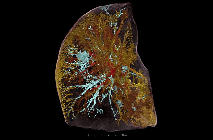
HiP-CT image of a whole left lung lobe from a 54-year-old male donor showing injury due to COVID-19. The lung is severely damaged. Aerated air spaces have been colored in cyan. There are areas where the airways are blocked, and the shape of the bunches of alveoli is very altered in comparison with that of a healthy lung. Blocked blood vessels are shown in yellow, and open blood vessels are shown in red.
There are few synchrotron radiation facilities, so this technology is not widely available. Because of the very high radiation dose, the technique will be used ex vivo for the foreseeable future, Walsh said.
"The x-ray dose is incredibly high; 2-kg normal CT scans are approximately 100 mG [milligauss]. This is 20,000 times more than a medical CT scan," explained Walsh. "We don't really have plans for this to become an in vivo human technique. We are aiming that we will be able to register clinical scans to HiP-CT in a few cases, and so HiP-CT will become a calibration for analyzing clinical techniques."
Elsie T. Nguyen, MD, FRCPC, vice-president of the Canadian Society of Thoracic Radiology and associate professor of radiology, University of Toronto, Toronto, Canada, noted that the technology will be valuable in pathology and radiology.
"HiP-CT appears to be an exciting new development that can help physicians, including radiologists, understand pathology that was once beyond the spatial resolution of computed tomography scans," said Nguyen in an interview with Medscape. "The fact that vascular abnormalities particularly relating to severe COVID-19 pneumonia can be visualized to the micron level is very novel and exciting. This will help us understand better from a mechanistic point of view what is happening to the blood vessels that contributes to worse outcomes, like shunting of blood or blood clots, and may have applications for prognostication to predict which patients are likely to survive severe COVID-19 pneumonia."
Nguyen noted that HiP-CT could help thoracic radiologists better visualize honeycomb cysts associated with fibrotic interstitial lung disease (ILD). It could help to classify the type of fibrotic ILD and inform patient prognosis.
"Currently, we struggle to differentiate early honeycomb cysts, which are a sign of more advanced lung destruction, from traction bronchiolectasis, that is, dilated airways due to surrounding fibrotic lung, on high-resolution computed tomography of the lungs," said Nguyen. She said HiP-CT was very promising and had many applications in addition to visualizing the lungs.
The research was funded by the Chan Zuckerberg Initiative, the ESRF, the UK-MRC, and the Royal Academy of Engineering. Walsh and Nguyen have disclosed no relevant financial relationships.
Nat Methods. Published online November 4, 2021. Full text


Commentaires
Enregistrer un commentaire